Med. Biol. Eng. Comput. 47(7) 719-729, July 2009.
links
full text from the publisher (open access)
abstract
The boundary-element method (BEM) is widely used for electrocardiogram (ECG) simulation. Its major disadvantage is its perceived inability to deal with the anisotropic electric conductivity of the myocardial interstitium, which led researchers to represent only intracellular anisotropy or neglect anisotropy altogether. We computed ECGs with a BEM model based on dipole sources that accounted for a "compound" anisotropy ratio. The ECGs were compared with those computed by a finite-difference model, in which intracellular and interstitial anisotropy could be represented without compromise. For a given set of conductivities, we always found a compound anisotropy value that led to acceptable differences between BEM and finite-difference results. In contrast, a fully isotropic model produced unacceptably large differences. A model that accounted only for intracellular anisotropy showed intermediate performance. We conclude that using a compound anisotropy ratio allows BEM-based ECG models to more accurately represent both anisotropies.
lay abstract
The beating heart generates small electric currents that flow through the body. The origin of these currents lies in processes at the level of the cell membrane. The currents serve to synchronize the contraction of the heart's two billion muscle cells, which makes the heart such an efficient blood pump. A side-effect of these currents is that they generate small electric potentials at the body surface which we can measure as an electrocardiogram (ECG). The ECG is an important diagnostic tool for cardiologists. Because it is relatively cheap and fast, and provides information about almost everything that happens in the heart, it is one of the first tests applied to any patient.
Most of our knowledge of the ECG is based on experience. For a real understanding of the relation between disease and ECG changes, especially if the changes are subtle, it is useful to simulate ECGs with a computer model. A hypothetical disease mechanism can be put into a model, which can then compute the ECG changes that would result from it. These can be compared to the ECG changes seen in real patients, to learn about what is actually going on in these patients.
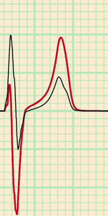
This paper makes two important but rather technical points about methods used for computer modeling of the ECG. First, it shows very accurately how important the anisotropy of the heart muscle is. Anisotropy refers here to the fact that electricity flows more easily along than across cardiac muscle fibers. The conclusion here is that models that do not account for this anisotropy give unreliable estimates of the ECGs measured on the chest, while they are still quite reliable for those measured from the arms and legs. The second point is that a relatively simple technique for ECG simulation, the boundary-element method, can be adapted to account for this anisotropy. This means that ordinary PCs can be used for reliable ECG simulation, rather than supercomputers.
erratum
The power 3 appearing in a numerator in equation (7) should not be there. This error occurs in the publisher's PDF version.
funding
Computational resources for this work were provided by the
Réseau québécois de calcul de haute performance
(RQCHP).