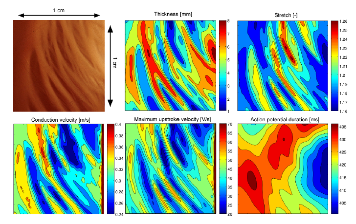
Effect of trabecular structure on impulse propagation in a 1-cm × 1-cm slab of atrial tissue. A planar depolarization wave was generated by stimulating the left side of the tissue slab. Overall stretch ratio was 1:2. Top: photographic picture of atrial tissue from rabbit [Eijsbouts et al 2003]; thickness as computed from the brightness of the picture; stretch ratio just before electrical stimulation. Bottom: conduction velocity; maximum upstroke velocity (dVmem/dt)max; action potential duration (APD at -60mV). The higher conduction velocity and (dVmem/dt)max on the right is a boundary effect.