other versions
abstract in J. Electrocardiol. 40 Suppl. (2007) page S49
conference proceedings paper in Anatol. J. Cardiol. 7 Suppl 1
(2007) pp 164-167 (poor graphics)
manuscript for conference paper (with better graphics)
context
This conference paper is the first demonstration in a
complete 3-D heart model that repolarization time, defined as the
instant of fastest downstroke of the action potential, coincides with
the instant of fastest upstroke of the local electrogram. It also
describes a preliminary version of a simple model for the local
electrogram, a rule-of-thumb model intended to help understanding
this relation intuitively. An improved version of the simple model was
published later in
Am. J. Physiol. H.
abstract
introduction
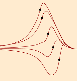
As a measure of repolarization time (Tr), the
instant of maximum slope (Tu) of the T wave in the local
unipolar electrocardiogram is commonly used. Although this method has
been well established both theoretically and experimentally, recent
observations on positive T waves in human hearts have
caused a renewed debate, involving also the theoretical basis for the
use of Tu. The purpose of this study was (1) to elucidate the
mechanism that leads to positive and negative T waves and
(2) to investigate theoretically which electrocardiogram feature best
predicts Tr.
methods
We used a bidomain reaction-diffusion model of the
human heart with anisotropic myocardium, transmural fiber rotation,
and heterogeneous ion-channel properties. This model calculates both
propagating action potentials (AP) and electrocardiograms. To explain
positive T waves, we compared results with those of a
much simpler model, which predicts T waves from local and
remote AP. We simulated normal tissue, repolarization abnormalities
and fibrotic tissue.
results
Repolarization time was defined as the instant of
steepest downstroke of the AP. The sign of the T wave was
almost uniquely determined by Tr. Positive T waves
occurred at early-repolarizing sites. In healthy tissue, the 2 models
agreed on T-wave sign in 92% of sites and predicted similar T
waves. This demonstrates that T-wave shape is determined primarily by
the difference between the local AP and the average AP in the
ventricles. Correlation between Tu and Tr was above 0.99 in both
negative and positive T waves.
conclusions
Our study predicts that (1) The sign of the T
wave is primarily determined by the difference between local AP
and the average AP in the ventricles; (2) positive T
waves occur at earlyrepolarizing sites; (3) Local Tr is best
estimated by Tu, also in positive T waves; and 4)
scarring and fibrosis may preclude any repolarization measurement.
funding
Computational resources for this work were provided by the
Réseau québécois de calcul de haute performance
(RQCHP). M. Potse was supported by a postdoctoral
research award from the Groupe de recherche en sciences et technologie
biomédicale (GRSTB), École Polytechnique and
Université de Montréal; and by the Research Center of
Sacré-Coeur Hospital, Montréal, Québec, Canada.
|