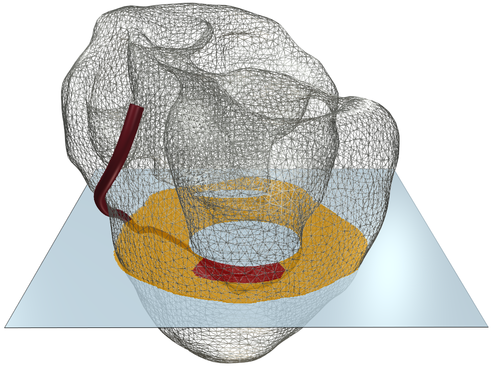
This figure shows how the diffusion of extracellular potassium in the heart affects potential pattern that would be expected in case a small subendocardial ischemic zone is present (red in the upper panel).
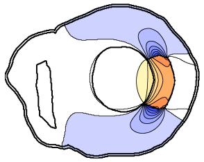
The lower left panel shows extracellular potentials in the case that this diffusion is absent. There is then an abrubt change in extracellular potassium concentration. The resulting gradient in membrane potential is very steep, even if somewhat smoothed due to "electrotonic" coupling between the cells. The extracellular potential pattern is quite unrealistic, with extrema (dark red, positive; and dark blue, negative) near the ends of the zone.
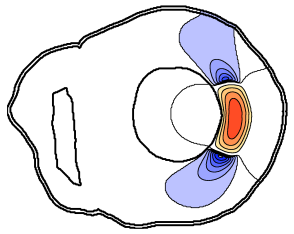
The lower right panel shows extracellular potentials in the case diffusion is present. A smooth gradient of potassium concentration and membrane potential occurs. Potassium and resting membrane potential are highest in the center of the ischemic zone, and so is the largest offset in extracellular potential (red).
Extracellular potentials in this figure are virtual ST-segment potentials. They are actually caused by depression of the TQ segment, because they are due to changes in resting membrane potential.